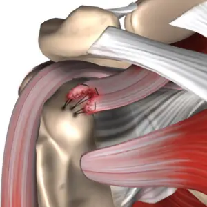
Orthopedic surgeons treat problems in your bones, joints, and muscles, and they diagnose joint pain and use several tools to find its source. When you feel pain in your knee, hip, or shoulder, they follow a clear process to understand it. Here is more information on the main steps they take to approach joint pain:
Physical Examination
A physical examination usually begins the diagnosis. The surgeon looks at the joint, and then presses on it to find tender spots. Since pain shifts during movement, they watch how the joint bends and rotates.
The orthopedic surgeon checks your range of motion. When swelling or stiffness appears, that detail guides the next step of the exam. They may ask you to walk, stand, or lift the limb, and they note where the pain starts.
Medical History Review
Your medical history gives the surgeon useful background. They ask about past injuries, and they note any surgeries you have had. Since family conditions like arthritis run in some families, the doctor asks about your relatives. During this review, the surgeon asks specific questions:
- When the pain began and how long it lasts
- Which activities make it worse
- How many medications you take now
- Which treatments you have tried
These answers narrow the possible causes. The surgeon writes down each detail, and the record helps track changes over time. When patterns emerge from your answers, the doctor forms an early idea of the problem.
Selective Imaging Techniques
Imaging shows what the surgeon cannot see from the outside. Since different problems need different views, the doctor picks the right scan for your case. X-rays reveal bone alignment, and they highlight fractures or worn cartilage.
MRI scans show soft tissue like ligaments and tendons. A CT scan gives detailed cross-sections, and it helps with complex bone shapes. When a joint needs a closer look at fluid or swelling, an ultrasound may be used.
X-rays, MRIs, CT scans, and ultrasounds each play a unique role in providing the most accurate picture of an injury or condition. Doctors rely on these tools to make informed decisions quickly. While X-rays are ideal for assessing bone injuries, an MRI is needed for spotting subtle issues in ligaments or other soft tissues that might not be visible through other methods.
Additional Lab Tests
Lab tests add another layer to the diagnosis. Blood work checks for infection, and it flags markers linked to inflammation. Since certain forms of arthritis may leave clues in your blood, these results help sort one condition from another. Sometimes the surgeon draws fluid from the joint, and this sample goes to a lab, where technicians examine it under a microscope. When crystals or bacteria appear in the fluid, the finding points to a specific cause.
Visit an Orthopedic Surgeon
Each step builds a fuller picture of your joint pain; the exam, history, imaging, and lab work fit together, and the surgeon uses them to name the cause. Since joint pain has many sources, this careful process matters for accurate results. Book an appointment with an orthopedic surgeon, and bring notes about your symptoms. When you take that first step, you give the doctor the details needed to start.
- Innovative Vein Treatment Techniques Revolutionizing Care
- The Potential Benefits of Stem Cell Therapy for Joint Pain Relief
- The Hyperbaric Chamber Treatment Process
- How Orthopedic Surgeons Approach Joint Pain Diagnosis
- Common Causes and Treatments for Erectile Dysfunction