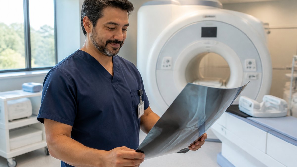
Musculoskeletal problems affect joints, muscles, tendons, and ligaments, and they often show up first as pain, stiffness, or limited motion. A medical diagnostic imaging center helps translate those symptoms into clear findings. While imaging does not replace a physical exam, it supplies visual evidence of what is happening beneath the skin.
Symptoms and Diagnosis
When a person reports knee pain after a fall or shoulder pain after repetitive overhead work, a medical diagnostic imaging center provides images. Standard X-rays show bone alignment and joint spacing, so they help with questions about fractures or advanced arthritis. Ultrasound and MRI add views of soft tissues. They help when a tendon, ligament, or muscle injury is suspected but not obvious on initial exam. The combination of history and targeted imaging gives the referring clinician a complete picture, and it supports planning for treatment.
Supports of Imaging Care
Diagnostic imaging feeds into each stage of musculoskeletal care. This changes the plan from the first assessment through return to activity. When a person presents with an injury, imaging helps identify what the problem involves. Follow-up imaging sometimes tracks healing, and it gives objective information about when a structure looks ready for progressive loading. Imaging also helps differentiate between persistent structural problems and symptoms driven by stiffness, weakness, or movement patterns. You may need multiple imaging tests done. The care team can adjust therapy or discuss other options.
It also supports targeted procedures around specific structures, because the clinician sees the needle path in real time. The practice notes that ultrasound imaging is used for musculoskeletal diagnosis, and it is used for treatment guidance. This reflects how dynamic imaging fits into modern orthopedic workflows. When imaging aligns with a detailed physical exam, it reduces uncertainty and supports tailored exercise, bracing, or procedural plans.
Types of Musculoskeletal Imaging
Different musculoskeletal problems call for different imaging tools, and a center uses a range of modalities. Since each modality has strengths, the referring clinician selects one. This is based on the area involved, the suspected injury, and whether bone or soft tissue needs evaluation.
- X-ray: Shows bones and joint alignment, so it helps detect fractures, dislocations, and some degenerative changes.
- Ultrasound: Uses sound waves to visualize tendons, ligaments, and muscles, and it also allows dynamic assessment during movement.
- MRI: Produces detailed images of soft tissues and bone marrow, and it helps with complex joint injuries and stress reactions.
- CT scan: Offers cross-sectional bone detail, so it supports evaluation of complex fractures and joint surfaces.
- Image-guided procedures: Use real-time imaging to guide needles for injections or aspirations around joints and soft tissues.
Book a Medical Diagnostic Imaging Center
People with joint, muscle, or tendon symptoms may benefit when clinical assessment is paired with focused diagnostic imaging. A diagnostic imaging center supplies the detailed views that help clinicians match treatment to the actual structure involved, and this supports clearer timelines and expectations. To learn more about musculoskeletal ultrasound and other diagnostic imaging options, contact your provider and ask how imaging fits into your current orthopedic or sports medicine plan.
- The Role of Testosterone Replacement in Treating Erectile Dysfunction
- The Long-term Benefits of Root Canal Therapy for Your Smile
- Identifying and Treating Common Warts
- Utilizing Telehealth for Accessible Chiropractic Consultations
- The Role of a Dermatologist in Managing Chronic Skin Conditions